How do I get started?
EXPERT ORTHODONTISTS
MANUFACTURED IN INDIA
CUTTING-EDGE TECHNOLOGY
ONTIME DELIVERY
We assist Orthodontists and Oral & Maxillofacial Surgeons with the following services which assist in performing the surgery:


We provide a post-surgery profile outcome prediction superimposed on the patient’s face along with values which are required at the time of the surgery.


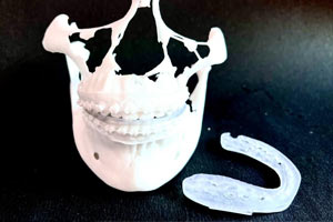
We provide auto-clavable high quality intermediate and final surgical splints which are made of biocompatible resin for medical grade applications.


We are also coming up with custom surgical guides which will further aid in precise surgical procedures.
The CT scan can be taken with a bite block, but the block should be thin, ensuring minimal distance between the jaws.
No orthodontic changes to be made after taking the digital scan/ impression. Either STL files can be uploaded, or you can courier the impressions/ stone models to us.
Pictures are required to undertake post-surgery profile outcome prediction. Extra-oral photographs should be taken with 1cm stickers on the temple & forehead.
Virtual Surgical Planning is designed to be time saving and efficient. Each virtual plan follows a similar workflow - structured and repeatable for both surgeon and staff allowing for quick turnaround.
Anatomical Assessments
We perform around 50 different types of 2D and 3D analysis as a part of pre-operative workflow and discuss the observations with you.
Virtual Planning and online discussion
Our experts conceptualize the surgical plan and discuss it online with the Surgeon and Orthodontist, making necessary modifications to the plan in real time.
3D printing
Once the plan is finalized, we 3d print the surgical splints, guides, and models according to your requirements.
Actual Surgery
We provide a case report which outlines values and step-by-step of the pre surgical plan, serving as a reference before surgery and in the operating theatre.

Expert Personnel: Our treatments are planned by expert Orthodontists and Surgeons who have over 2 decades of experience in planning and performing treatments.
Quick Turnaround: We deliver splints within 10 days of sharing treatment prerequisites. Also, in case of exigencies, we take cases up on priority basis.
Explicit communication: “The result of bad communication is a disconnection between strategy and execution.” We carefully discuss the plan and get approvals through video calls.
Cost Effective: Bundled services are supplied to patients at an affordable cost that considerably outweighs the advantages.
Multiple Treatment plans: We simulate distinct surgical procedures to obtain optimal results for the patient.
VSP is a disruptive innovation that eliminates the shortcomings of conventional planning methods and seeks to benefit multiple stakeholders.
VSP eliminates the need of performing pre-surgical procedures via traditional articulator method and facebow transfers, which has a high probability of errors. Also, since all the records are digitally available - both 2D and 3D, the orthodontist can plan the surgeries far quicker compared to traditional methods.
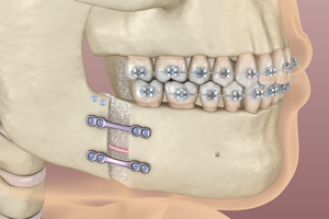
Three-dimensional imaging enables the surgeon to establish necessary osteotomy planes preoperatively and assess different surgical scenarios. Further, the time taken to perform a surgery based on a virtual plan is significantly lesser than under a conventional plan with the aid of surgical splints and guides.
VSP allows the patients to see the post-surgical profile prediction superimposed on their face prior to the surgery which allows them to decide whether to go ahead with the surgery, which was not possible earlier. Further, VSP aids quicker surgeries, reducing the time spent in the OT.
Medical grade CT – Full skull to C7. If Medical Grade CT is not available, CBCT of the entire skull to C7 will do. Due to the grainy nature of CBCTs however, there is possibility of severe distortion in the profile outcome prediction. Hence, CTs are recommended.
Partial CBCT (only dental arches) will not work.
Maxillary and Mandibular arch with the bite in STL Format. If an intra oral scan is not available, stone models with minimum air bubbles are required. The upper and lower impression should be sent along with a wax bite (in present occlusion).
Note – No orthodontic changes to be made after taking the scan/impression
Intra-oral and Extra oral photographs of the patient – Intra-oral photographs: Occlusal Frontal View, Right Molar Retention, Left Molar Retention, Occlusal View of Upper, Occlusal View of Lower
Extra-oral photographs: Primarily for generating post-surgery profile outcome prediction. The photographs should be taken with 1cm stickers on the temple & forehead
The CT can be taken with a bite block, but the block should be thin, ensuring minimal distance between the jaws.
Slice thickness: CT scan < 0.6 mm and CBCT 0.35 mm to 0.50 mm
Format: DICOM file
Pre-treatment COGS/Burstone Analysis (2D)
50 different types of 2D analysis
Arnett Analysis (3D)
Surgical movements (2D & 3D)
Post-surgery profile outcome prediction
Yes, we can provide only the Cephalometric / Pre-treatment COGS analysis if necessary. The prerequisite for it is only a Lateral Cephalogram.
Yes. If you already have the treatment plan ready, you just send us the STL file of the intermediate & final splints.
It is compulsory to send a dental scan /dental cast /impression due to the reduced quality of image of the dental surfaces obtained from tomography.
Note – No orthodontic changes to be made after taking the scan/impression
VPS gives three-dimensional imaging which enables the surgeon to establish necessary osteotomy planes preoperatively and assess different surgical scenarios.
It also provides a prediction of the post-surgery profile outcome which aids in effective communication between the surgeon and the patient. In the conventional method we cannot show the outcome of the surgery before the actual surgery is performed.
VSP also allows for minimal invasive surgeries as it follows the osteotomy line nearly the same as in a real intraoperative osteotomy.
No. The use of facebow transfer and expensive articulators are eliminated by VSP. The analysis and treatment plan for the surgery is prepared using a software. Once you finalize the treatment plan, the splints are 3D printed.
Learn how Route to Smile is helping doctors provide specialized care to their patients.