What are the pre-requisites for Virtual Surgical Planning?
What is Virtual surgical planning?
The
Virtual Surgical Planning (VSP) method for 3D planning and analysis has been
expanding of late. Virtual Surgical Planning consists of analyzing the
patient’s skeletal deformity with a 3D analysis program using CT, performing
virtual surgery, and then fabricating surgical splints using 3D printing
technology.
Virtual surgical
planning uses computer graphics to enhance the predictability of results,
standardize technique, and improve communication between the patient and the Dentist
(Orthodontist as well as the Oral & Maxillofacial Surgeon).
What are the pre-requisites for Virtual surgical planning for orthognathic surgery?
1. Patient's intraoral and extraoral photographs – This helps us assess the existing condition of the patient. It also ensures that we can provide a prediction of the likely post-surgery outcome of the patient’s profile where the patient can appreciate the changes in their soft tissue.
The soft tissue prediction will also help the doctor in better communication with the patient. The patient can be educated about the procedure that he/she will undergo and the patient will be motivated to go ahead with the procedure after seeing the predicted outcome.
2. Medical grade CT from glabella to C7 – If a medical grade CT is not available, CBCT will do. The CT can be taken with a bite block, but the block should be thin, ensuring minimal distance between the jaws.
Properties of CT/CBCT –
- Partial
CBCT (only dental arches) will not work
- The
slice thickness for a CT scan should be less than 0.6 mm
- The
slice thickness for a CBCT should be between 0.35 mm to 0.5 mm
- CBCTs
should not be taken with a chin support
- CT/ CBCT data should be provided in DICOM format
- It must be noted that due to the grainy nature of CBCTs there is a possibility of severe distortion in the profile outcome prediction. Hence CTs are recommended.
With the CT/ CBCT data, we can generate the lateral cephalogram and OPG of the patient making it easier for the Doctor to prepare the treatment plan.
A major contribution provided by CT images and planning
software is a more accurate evaluation of the subject’s airway. Maxilla and
mandible movements significantly influence the airway volume of the subject.
Therefore, the airway should be considered in this phase of planning the
treatment.
3. Maxillary and mandibular arch with the bite in STL format/ Impression – Due to the metal brackets, there is distortion in the in maxillary and mandibular arch in a CT /CBCT scan. To get a clear picture of the dental arch, we need to take an impression of the patient or an intra oral scan.
You must make sure that there are no orthodontic changes made after taking the scan /impression.
In case you are taking an intra-oral scan, the STL file will be directly generated.
In the case of stone models, the STL files can be generated using a desktop scanner. The impression should have minimum air bubbles. The upper and lower impression should also include a wax bite (in present occlusion).
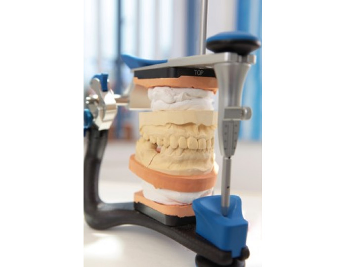
When we receive the above data, we can move forward with the case and prepare a precise virtual surgical plan without the need for expensive articulators, facebow transfers, mounting stone models or fabrication of manual splints.
Once you approve the treatment plan, the splints will be 3D printed and sent to you before the surgery date.
It’s time to Think Ahead. Think Virtual Surgical Planning.
Related Post

What are the benefits of Virtual Surgical Planning?
Virtual surgical
planning (VSP) has evolved into a useful tool for many surgeries.